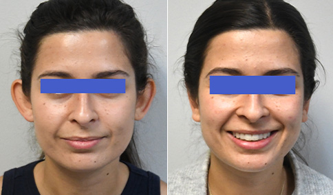
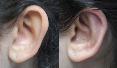
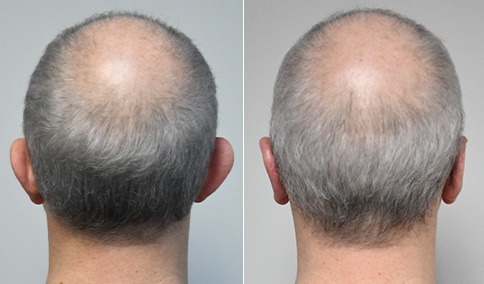
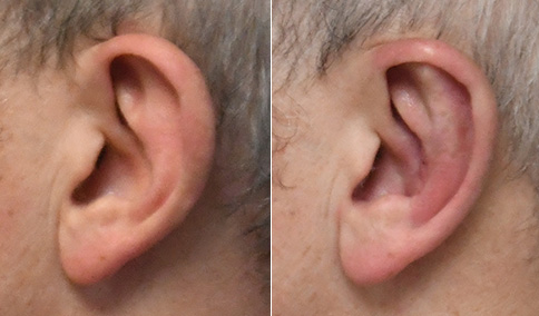
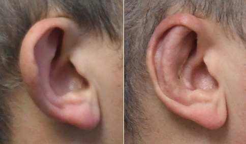
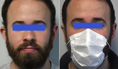
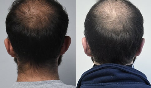
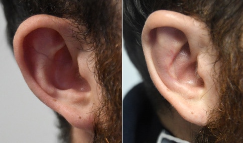
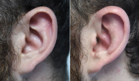
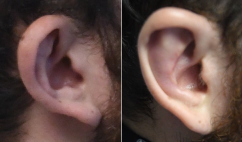
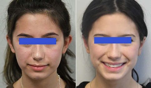
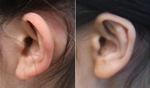
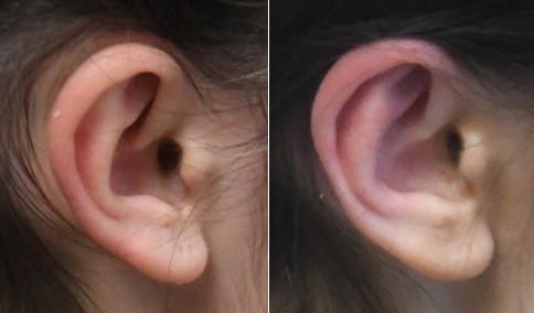
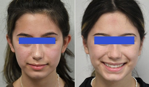
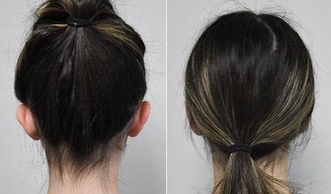
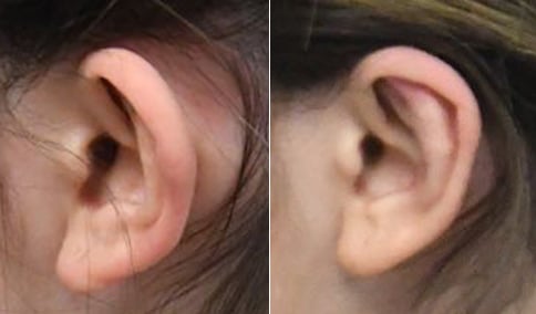
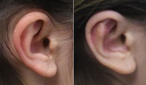
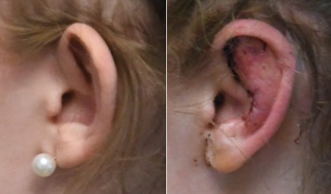
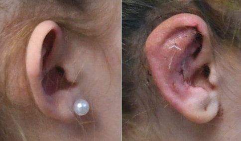
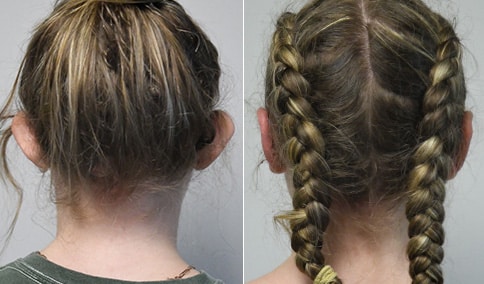
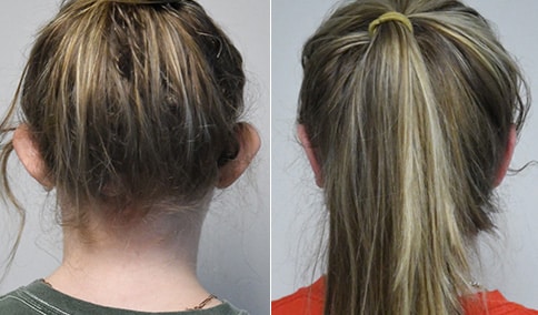
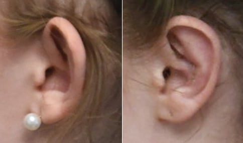
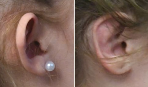
A woman in her late 20s who has been bothered by prominent ears her entire life. She is shown before and again, just 6 weeks after correction in the operating room.
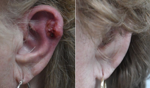
She still has some mild redness of the ears at 6 weeks, which should now fade and resolve over the next month. Scars are at their reddest and thickest at 6 weeks and then will fade and improve over the next 2 years. At 6 weeks, the incisions just inside the conchal rim are already starting to fade. During surgery, I first est back her deep concha by excision of a crescent of cartilage through an anterior approach. Then I rasped her antehelical fold with the Dingman otoabrader, as taught to me by Dr.Reed Dingman at the University of Michigan during my plastic surgery residency, to break the “spring” of the cartilage and allow it to bend with less force. Then, “Mustarde” sutures were placed to recreate the antithetical fold and give it a gentle curve. At this point, her ear lobes will still be prominent and were set back by dividing a ligament called the “intertragicohelcine ligament” and then pexing the helical tail to the concha with a suture as described by Dr. Richard Webster, along with a “fishtail” excision of skin behind the earlobe, without interfering with her ear piercing holes. She loves the natural appearing contour of her ears after surgery. She told me that the wind recently blew her hair up, and ordinarily, she would have been quite anxious that her formerly prominent ears would show, but she found that she was no longer bothered because of the now normal and beautiful contour.


Cosmetic & Plastic Surgery Specialist
"I treat my patients like I would treat
- Jonathan D. Hall, MD, FACSmembers of my own family."
Schedule Consultation